
Madison Ortho Knowledge Center

Orthopedic Surgical Insights & Innovations

Exploring clinical developments, advanced implant technologies, trauma fixation solutions, and news from the global orthopedic community.

0%

Trochanteric Reconstruction Nails are specialized intramedullary implants for difficult fractures around the hip and upper femur. They combine a strong central nail with proximal screws or blades. These components support the femoral neck, head, and trochanteric region from inside the bone.

Orthopedic trauma surgeon Dr. Kenneth J. Koval has emphasized a practical principle: “The implant is a tool; the treatment is the patient.” That idea matters here. A nail may improve stability, but it cannot replace accurate fracture assessment, careful reduction, or appropriate rehabilitation. In the operating room, surgeons may evaluate bone quality, fracture extension, limb alignment, and rotational control before selecting the implant. Small details matter. A few millimeters of malalignment can affect walking comfort and hip mechanics.

This guide explains what Trochanteric Reconstruction Nails are, how they work, and when surgeons may consider them. It also examines their construction, fixation options, potential advantages, and limitations. Readers will see how these nails differ from standard cephalomedullary devices and plates. The discussion will remain clinically grounded, while recognizing that implant choice depends on imaging, fracture pattern, patient health, and surgeon experience.

Not every complex fracture needs this nail. That is worth remembering. Evidence and surgical judgment must guide the decision. Even a technically successful fixation can face challenges, including delayed healing, screw migration, infection, or reduced mobility. Better understanding begins with realistic expectations, not promises.
What Are Trochanteric Reconstruction Nails?

Definition and Purpose of Trochanteric Reconstruction Nails

Trochanteric reconstruction nails are metal intramedullary implants designed for complex fractures near the upper femur.

They pass through the femoral canal and provide internal support along the bone’s length. Special proximal locking options help secure the femoral neck, head, or trochanteric region. Their definition is simple: they are load-sharing devices for difficult proximal femur injuries.

Their purpose is to restore stability while allowing controlled movement during healing.

Surgeons may consider them for fractures extending from the greater trochanter toward the shaft or femoral neck. The exact design and screw position depend on fracture lines, bone quality, and imaging findings. The choice is not always obvious. A nail can stabilize bone, but it cannot replace accurate reduction, careful surgery, or rehabilitation. Even experienced teams must balance fixation strength with soft-tissue protection.

Tips: Ask whether the fracture reaches the femoral neck or shaft. Request a clear explanation of the planned fixation points. Follow weight-bearing instructions closely. Report increasing pain, swelling, fever, or wound drainage promptly. Recovery may feel uneven, and that deserves discussion. A thoughtful plan can change as new images, symptoms, or healing patterns appear.

Anatomy and Fracture Types They Are Designed to Treat

What Are Trochanteric Reconstruction Nails?

Anatomy and Fracture Types They Are Designed to Treat

Trochanteric reconstruction nails are intramedullary implants placed inside the femoral canal. They support fractures near the greater and lesser trochanters. These regions transfer body weight from the hip joint into the femoral shaft. Their anatomy is complex. Muscle forces can pull fragments upward, outward, or into rotation.

These nails are commonly designed for unstable intertrochanteric fractures, subtrochanteric fractures, and reverse-obliquity patterns. They may also support fractures extending from the trochanteric region into the femoral neck or shaft. Proximal screws or blades help control the femoral head and neck. Distal locking helps limit rotation and shortening. Shorter is not always better. Nail length, canal shape, fracture extension, and bone quality all influence selection.

The International Osteoporosis Foundation reports approximately 1.6 million hip fractures worldwide each year. Its projections suggest this number could reach 2.6 million by 2025 and 4.5 million by 2050. This rising burden increases the need for stable fixation, especially in fragile bone. The American Academy of Orthopaedic Surgeons’ 2021 guideline supports cephalomedullary fixation for subtrochanteric fractures and selected unstable intertrochanteric injuries. However, evidence is not perfectly uniform across every fracture pattern. A nail cannot correct poor reduction. Careful imaging, soft-tissue assessment, and surgical experience remain essential.

Key Components and Structural Features

What Are Trochanteric Reconstruction Nails?

Key Components and Structural Features

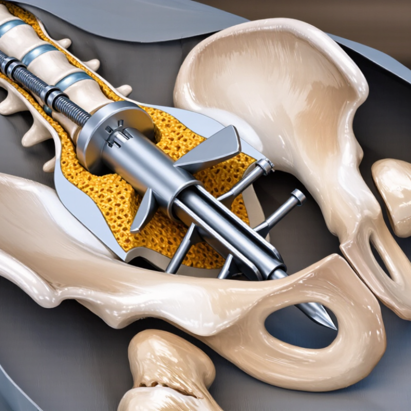
Trochanteric reconstruction nails are intramedullary implants designed to stabilize fractures near the greater trochanter, femoral neck, and subtrochanteric region. They sit inside the femoral canal, sharing load with the bone rather than resting entirely on its surface. Their shape usually includes a gentle curvature that follows the femur’s natural alignment.

The proximal section contains carefully positioned openings for reconstruction screws or blades. These components pass into the femoral head and neck, helping control rotation and maintain fracture alignment. Some designs use two proximal screws to improve rotational stability. The distal part includes locking holes, which limit shortening and unwanted movement. Small details matter.

An end cap can close the nail and support later removal when clinically necessary. A targeting guide helps the surgeon place screws through the nail with greater accuracy. During treatment, fluoroscopic imaging confirms entry point, depth, alignment, and screw position. That process requires judgment, not just instrument handling.

In practical use, nail diameter, length, curvature, and screw configuration must match the patient’s anatomy and fracture pattern. Poor reduction can remain a problem, even with a strong implant. This is easy to underestimate. Bone quality, soft-tissue damage, and the stability of the lateral wall also influence the result. Clinical decisions should follow current orthopedic evidence, careful imaging, and the treating surgeon’s experience.

Surgical Placement and Fracture Stabilization Process

What Are Trochanteric Reconstruction Nails?

Surgical Placement and Fracture Stabilization Process

Trochanteric reconstruction nails are intramedullary devices used to stabilize complex fractures near the hip. They may support breaks involving the greater trochanter, femoral neck, or upper femoral shaft. Their design allows controlled fixation through the main canal and the proximal femur.

Careful planning begins with radiographs and, when necessary, computed tomography scans. The surgical team studies fracture lines, bone quality, and the patient’s overall condition. During surgery, the patient is positioned to allow traction and imaging. The surgeon reduces the fracture, then introduces a guidewire through an entry point near the greater trochanter. Reaming may prepare the canal for the nail. Fluoroscopy helps confirm alignment throughout the procedure.

After nail insertion, proximal screws or blades cross into the femoral head and neck. These components resist rotation and collapse. Distal locking screws improve control of length and rotation. The fixation must match the fracture pattern, not simply follow a routine sequence. Small alignment errors can affect walking and healing.

The process is precise, but rarely perfect. Osteoporotic bone, swelling, or unstable fragments can complicate reduction. A screw may appear well positioned on one view yet require correction on another. This is why repeated imaging and intraoperative judgment matter. Postoperative weight bearing depends on fixation strength, fracture stability, bone health, and the surgeon’s assessment. Recovery also requires monitoring for infection, implant failure, delayed healing, and reduced hip movement.

Benefits, Risks, and Postoperative Considerations

What Are Trochanteric Reconstruction Nails?

Trochanteric reconstruction nails are internal devices used to stabilize complex fractures near the upper femur. They can support fractures extending through the trochanteric region and femoral neck. Their design may reduce movement between broken bone fragments. This stability can help patients begin controlled mobility earlier, when medically appropriate. Surgical planning still depends on fracture pattern, bone quality, age, and general health.

Benefits, Risks, and Postoperative Considerations

A major benefit is load sharing along the femur, which may support alignment during healing. Smaller incisions may also reduce soft-tissue disruption compared with some open procedures. However, risks include infection, blood clots, bleeding, nerve injury, malalignment, delayed healing, and implant failure. Pain near the hip or thigh can persist. Even careful planning has limits. A stable fixation does not guarantee painless movement.

After surgery, weight-bearing must follow the surgeon’s instructions. Some patients use a walker for several weeks. Physical therapy usually targets ankle motion, quadriceps strength, balance, and safe transfers. Patients should watch for increasing redness, wound drainage, fever, calf swelling, chest pain, or sudden shortness of breath. These symptoms need urgent medical assessment. Follow-up X-rays help evaluate alignment and bone healing. Recovery is rarely linear. Weakness may improve slowly, and the plan may change when imaging or function reveals an unexpected problem. Nutritional support, smoking cessation, and prescribed medication use can also influence healing.

FAQS

What are trochanteric reconstruction nails?

They are metal intramedullary implants for complex fractures near the upper femur. They sit inside the femoral canal and share load with the bone.

What injuries may require this type of nail?

Surgeons may consider them when a fracture extends from the greater trochanter toward the shaft or femoral neck. Imaging helps define the fracture path.

How does the nail support fracture healing?

The nail restores internal stability while allowing controlled movement. Proximal screws can support the femoral neck, head, or trochanteric region.

What are proximal and distal locking options?

Proximal openings hold reconstruction screws or blades near the femoral head and neck. Distal locking holes reduce unwanted shortening and movement.

Why might two proximal screws be used?

Two screws may improve rotational stability and help maintain alignment. Their use depends on the fracture pattern and bone quality.

How does the surgical team check screw placement?

Fluoroscopic imaging checks the entry point, depth, alignment, and screw position. Small positioning errors can matter.

What factors influence nail selection?

Nail diameter, length, curvature, and screw configuration should match the patient’s anatomy. Bone quality and lateral-wall stability also influence planning.

Can a strong nail correct every fracture problem?

No. The implant cannot replace accurate reduction, careful surgery, or rehabilitation. A strong device may still perform poorly after poor reduction.

What should patients monitor during recovery?

Follow weight-bearing instructions closely. Report increasing pain, swelling, fever, or wound drainage promptly.

Is recovery always steady?

Not always. Progress may feel uneven, and new images or symptoms may change the plan. That uncertainty deserves a clear discussion.

Conclusion

Trochanteric Reconstruction Nails are specialized orthopedic implants used to stabilize complex fractures around the upper femur, particularly injuries involving the trochanteric region and extending toward the femoral shaft. Their purpose is to restore alignment, maintain bone length and rotation, and support healing while allowing controlled movement. These fractures may result from falls, high-impact trauma, or weakened bone, and treatment depends on the fracture pattern, bone quality, and the patient’s overall health.

The nail is inserted into the femoral canal and typically includes locking options, proximal fixation elements, and structural features designed to control fragments near the hip. During surgery, the fracture is reduced, the nail is positioned, and screws or related fixation components secure the trochanteric and shaft fragments. Potential benefits include strong internal support, improved alignment, and earlier mobilization. However, risks may include infection, blood loss, implant migration, delayed healing, malalignment, or the need for additional surgery. Postoperative care commonly involves pain management, imaging follow-up, physical therapy, and gradually increasing weight-bearing according to healing progress.

Olivia

Olivia

Olivia is a dedicated marketing professional with a deep expertise in the company's product offerings. Her passion for innovative solutions and market trends allows her to craft compelling narratives that resonate with customers and stakeholders alike. Olivia regularly updates the company's......